
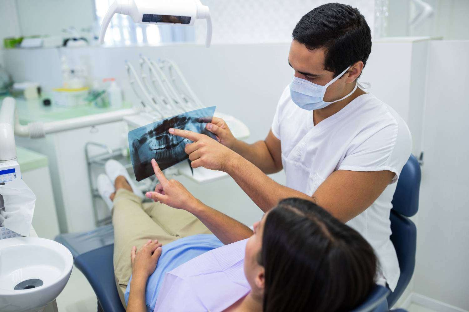
A dental x-ray lets your dentist see what a mirror and a light cannot: decay between teeth, infection at the tip of a root, bone levels around the teeth and wisdom teeth still hidden in the jaw. For many patients, however, the first question is whether the radiation is safe.
This guide explains the main types of dental X-rays, what each one shows, how much radiation they involve and how your dentist decides when you actually need one.
Why do dentists take a dental x-ray?
A clinical examination shows only the visible surfaces of your teeth and gums. In contrast, X-rays reveal what lies between and beneath them. Your dentist may use them to:
- Find cavities between teeth and under old fillings before they become painful
- Check for infection or abscesses at the root tips
- Measure bone loss caused by gum disease
- See impacted or unerupted teeth, including wisdom teeth
- Plan treatments such as root canals, implants, extractions and braces
- Monitor how a child’s teeth and jaws are developing
In short, X-rays help your dentist catch problems early, when treatment is usually simpler.
Types of dental x-ray
Dental X-rays fall into two broad groups. Intraoral X-rays use a small sensor placed inside the mouth and show a few teeth in fine detail. Extraoral X-rays, on the other hand, capture the whole mouth, jaws or skull from outside.
Bitewings
You bite gently on a small tab while the sensor records the crowns of your upper and lower back teeth together. Bitewings are especially good at finding decay between back teeth and checking the bone level along the gums.
Periapical view (IOPA)
An intraoral periapical X-ray, often called an IOPA in India, shows one or two teeth from the crown to the tip of the root and the bone around it. Dentists rely on it to diagnose abscesses, cracked roots and to guide root canal treatment.
Occlusal view
This view captures the floor or roof of the mouth in one image. Dentists use it mainly in children to track developing teeth, or to locate extra or blocked teeth.
Panoramic view (OPG)
An OPG, or orthopantomogram, gives a single wide image of all the teeth, both jaws and the jaw joints. The machine rotates around your head while you stand still for a few seconds. Dentists commonly use it to assess wisdom teeth, plan orthodontic treatment and get an overview before major work.

Lateral cephalogram
This side view of the skull shows how the jaws and teeth relate to each other. Orthodontists use it to plan braces and aligner treatment and to measure jaw growth.
CBCT scan (3D dental imaging)
Cone beam computed tomography, or CBCT, produces a three-dimensional image of the teeth, bone, nerves and sinuses. It is the preferred tool for planning dental implants, assessing complex root canals and locating impacted teeth close to nerves. Because a CBCT involves more radiation than a standard dental x-ray, dentists reserve it for cases where 3D detail genuinely changes the treatment plan.
Which dental x-ray is used for what?
| X-ray type | What it shows | Commonly used for |
|---|---|---|
| Bitewing | Crowns of upper and lower back teeth | Decay between teeth, bone level checks |
| Periapical (IOPA) | Whole tooth from crown to root tip | Abscesses, root canal treatment, cracked roots |
| Occlusal | Floor or roof of the mouth | Children’s developing teeth, blocked teeth |
| Panoramic (OPG) | All teeth, both jaws and jaw joints | Wisdom teeth, orthodontic planning, overall assessment |
| Lateral cephalogram | Side view of skull and jaws | Orthodontic planning |
| CBCT (3D) | Teeth, bone, nerves and sinuses in 3D | Implant planning, complex root canals, impacted teeth |
Is a dental x-ray safe?
Yes. Dental X-rays use very low doses of radiation. According to the American Dental Association, they expose patients to even less radiation than people receive from their everyday environment. Moreover, modern digital sensors need far less radiation than old film, and they display the image instantly. You can read more about this shift in our article on digital dentistry.
Even so, any radiation should be justified. For this reason, good practice is to take an X-ray only when it will help with a diagnosis or treatment decision, to use the smallest area and lowest dose that gives a clear image, and to avoid repeating images unnecessarily. In India, the Atomic Energy Regulatory Board (AERB) regulates the X-ray equipment that clinics use.
Do you need a lead apron?
Many patients remember wearing a heavy lead apron and thyroid collar. However, the American Dental Association updated its guidance in 2024 and no longer recommends routine lead aprons or thyroid collars for dental imaging, because modern equipment limits the beam to the area being imaged. Some clinics still offer one, and you are always welcome to ask.
Imaging during pregnancy
The ADA and the American Academy of Oral and Maxillofacial Radiology consider dental X-rays safe during pregnancy, since the dose is very low and the beam is aimed at the mouth. Nevertheless, always tell your dentist if you are pregnant or might be. They may postpone routine X-rays until after delivery and take only those needed for urgent care, such as a painful infection. Our guide to dental care in pregnancy covers other precautions.
How often do you need a dental x-ray?
There is no fixed schedule that suits everyone. Instead, your dentist decides based on your age, your history of decay and gum disease, and what they see during the examination. As a general guide:
- People with a low risk of decay and healthy gums may need bitewings only every few years
- People with frequent cavities, gum disease or extensive dental work usually need them more often
- Children often need X-rays more frequently than adults, because their teeth change quickly and decay spreads faster in baby teeth
- New patients may need a baseline set, unless recent X-rays are available from a previous dentist
Regular dental check-ups give your dentist the chance to decide when an X-ray is genuinely useful.
What to expect during a dental x-ray
- Your dentist or assistant explains which images they need and why
- You remove glasses, earrings, removable dentures or anything metal near the head
- For intraoral images, you bite gently on a small sensor holder for a few seconds
- For an OPG or CBCT, you stand or sit still while the machine rotates around you
- The images appear on screen almost immediately, and your dentist can talk you through them
The whole process is painless. If you gag easily, let the team know, since smaller sensors and a few simple techniques can make intraoral images more comfortable.
Can you get copies of your images?
Yes. Your X-rays belong to your dental records, and you can ask for a digital copy. This is especially helpful if you change dentists, seek a second opinion or travel for treatment, because it can avoid repeating the same images.
Digital imaging at Amaya Dental in Bengaluru
At Amaya Dental, we take X-rays only when they will guide your diagnosis or treatment, and we always explain what the images show. Digital imaging lets us show you your own teeth on screen, so you can see exactly why we recommend a treatment.
If you have a dental concern or are due for a check-up, book a consultation or contact our team.
The bottom line
A dental x-ray is a quick, low-dose way to find problems that an examination alone would miss. The right type and frequency depend on your mouth and your risk, so your dentist should explain why each image is needed. For more on radiation safety, see the ADA overview of dental X-ray safety.
Frequently Asked Questions About Dental X-Rays
Very little. Standard dental X-rays involve a very low dose, less than the background radiation people receive from their everyday environment. A CBCT scan involves more, which is why dentists use it only when 3D detail is necessary.
An OPG is a panoramic X-ray that shows all the teeth and both jaws in one image. An IOPA shows one or two teeth in fine detail, from the crown to the root tip.
Dental X-rays are considered safe during pregnancy because the dose is very low and the beam is aimed at the mouth. Still, tell your dentist you are pregnant, so they can postpone routine images and take only those needed for urgent treatment.
Often, yes. Children’s teeth and jaws change quickly, and decay can spread fast in baby teeth. Your dentist chooses the type and frequency based on your child’s age and risk.
A CBCT scan shows the height, width and density of the jawbone and the position of nerves and sinuses in 3D. As a result, the dentist can plan the implant’s exact position safely.
Yes, it is your choice. However, without X-rays your dentist may miss decay between teeth, infections or bone problems. Ask your dentist to explain what they are looking for, so you can make an informed decision.


